
Research Director: Dr. Setsuo Hirohashi
(Deputy Director, National Cancer Center Research Institute)
Research Term: 1993~1998
Research focused on the shapes ofcells as expressions of their genetics and the influences of the surrounding cells and inter-cellular environment while hoping to understand why cells have their particular shapes and functions and why they are built up with particular molecules. Research results have contributed to a most useful fusion similar to a Renaissance in morphology or pathology, which could be called “molecular pathology” or “molecular morphology.” This new discipline should contribute greatly to the clarification of the pathogenesis of various diseases, including cancer.
Research Results
New method for the reconstitution of three-dimensional images of non-stained living cells or tissues: The principle of computed tomography (CT), a well-known X-ray diagnostic technique, was applied to optical microscopy by developing a scanning optical CT microscope. This method has shown clear three-dimensional recon-structed images of living cells.
New method for efficient gene screening based on the expression pattern: High-speed performance in situ hybridization methods coupled with many sliced tissue preparations were developed and applied to isolate genes from various tissues based on their expression patterns.
New method for efrlCient gene screening based on the intracellular localization of products: A cDNA library in which truncated polypeptide fused with GFP (Green Fluorescent Protein) was constructed and expressed in cell lines with distinct polarity. By using signals from GFP as markers, many genes showing interesting intracellular localization were isolated.
Molecular cloning of a novel family of cadherin (a group of Calcium-dependent cell adhesion molecules) and analysis of the molecular dynamics of adhesion-dependent cadherin homodimer: Considering the diverse functions of cadherin in the regulation for mammalian cell adhesion and its disorder, three new members in humans were isolated to analyze its function in normal physiology and cancer metastasis. To explain cadherin-mediated cell adhesion, a chemical crosslinking analysis was introduced to the cadherin molecules expressed on the cell surface, A significant correlation was identified between the cadherin dimerization and adhesion activity in vivo.
Characterization of the three-dimensional reconstituted system of cancer tissue: Several cancerous cell lines embedded in collagen gel as a clump showed different morphologies and various degrees of mobility in response to externally added growth factors. A significant correlation of these in vivo observations to their metastic activity in vivo was confirmed. Genes responsible for these correlations were screened.
Isolation of genes involved in mesenchymal-epithelial interactions: Molecular analysis on chick digestive-tissue development allowed the isolation of several genes involved in this organ’s differentiation and morphogenesis. It was demonstrated that they are regulated by mesenchymal-epithelial interactions.
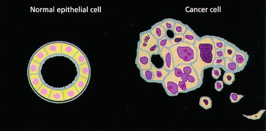
·Apparent differences of the cell adhesion and the cell polarity are shown between a normal epithelial cell and a cancer cell.
