The joint development team of Professor Shibata (the University of Tokyo), JEOL Ltd. and Monash University succeeded in directly observing an atomic magnetic field, the origin of magnets (magnetic force), for the first time in the world. The observation was conducted using the newly developed Magnetic-field-free Atomic-Resolution STEM (MARS)(1). This team had already succeeded in observing the electric field inside atoms for the first time in 2012. However, since the magnetic fields in atoms are extremely weak compared with electric fields, the technology to observe the magnetic fields had been unexplored since the development of electron microscopes. This is an epoch-making achievement that will rewrite the history of microscope development.
Electron microscopes have the highest spatial resolution among all currently used microscopes. However, in order to achieve ultra-high resolution so that atoms can be observed directly, we have to observe the sample by placing it in an extremely strong lens magnetic field. Therefore, atomic observation of magnetic materials that are strongly affected by the lens magnetic field such as magnets and steels had been impossible for many years. For this difficult problem, the team succeeded in developing a lens that has a completely new structure in 2019. Using this new lens, the team realized atomic observation of magnetic materials, which is not affected by the lens magnetic field. The team’s next goal was to observe the magnetic fields of atoms, which are the origin of magnets (magnetic force), and they continued technological development to achieve the goal.
This time, the joint development team took on the challenge of observing the magnetic fields of iron (Fe) atoms in a hematite crystal (α-Fe2O3) by loading MARS with a newly developed high-sensitivity high-speed detector, and further using computer image processing technology. To observe the magnetic fields, they used the Differential Phase Contrast (DPC) method (2) at atomic resolution, which is an ultrahigh-resolution local electromagnetic field measurement method using a scanning transmission electron microscope (STEM) (3), developed by Professor Shibata et al. The results directly demonstrated that iron atoms themselves are small magnets (atomic magnet). The results also clarified the origin of magnetism (antiferromagnetism (4)) exhibited by hematite at the atomic level.
From the present research results, the observation on atomic magnetic field was demonstrated, and a method for observation of atomic magnetic fields was established. This method is expected to become a new measuring method in the future that will lead the research and development of various magnetic materials and devices such as magnets, steels, magnetic devices, magnetic memory, magnetic semiconductors, spintronics and topological materials.
This research was conducted by the joint development team of Professor Naoya Shibata (Director of the Institute of Engineering Innovation, School of Engineering, the University of Tokyo) and Dr. Yuji Kohno et al. (Specialists of JEOL Ltd.) in collaboration with Monash University, Australia, under the Advanced Measurement and Analysis Systems Development (SENTAN), Japan Science and Technology Agency (JST).
<Terminology>
(1) Magnetic-field-free Atomic-Resolution STEM (MARS)
An electron microscope is an instrument to directly observe the microstructure in a sample, where an electron beam is injected into the sample, and the electron beams transmitted and scattered by the sample are magnified using a magnetic field lens. Currently, it is possible to directly observe atoms using an electron microscope. In an optical microscope, the spatial resolution is in principle limited to about one micrometer due to the light source (visible light). On the other hand, electron microscope is an instrument where this spatial resolution limit is exceeded by utilizing the wave nature of electrons. Therefore, it can be said that an electron microscope is an observation technology that applies the benefits of quantum mechanics in the most direct way. The Magnetic-field-free Atomic-Resolution STEM (MARS) is an electron microscope developed by the present joint development team in 2019, capable of measuring a sample in a magnetic-field free environment. For details, please refer to the following press release.
“An innovative electron microscope overturning common knowledge of 88 years history”(May 24, 2019)
https://www.jst.go.jp/pr/announce/20190524/index_e.html
(2) Differential Phase Contrast (DPC) method
A method to measure the electromagnetic field at each point in a sample. Specifically, when an electron beam is injected in a sample, the force of the electromagnetic field that exists within the sample causes a slight trajectory change in the electron beam incident, and by measuring the difference in the electron beam intensity detected in each position of a split detector, the electromagnetic field can be measured. Since the spatial resolution of this method is basically determined by the size of the electron probe, observation of an electromagnetic field at atomic resolution is in principle possible using the DPC method.
(3) Scanning Transmission Electron Microscope (STEM)
An instrument to directly observe the structure inside a sample. Specifically, a micro-focused electron beam is scanned on the sample, and observation is conducted by measuring the intensity of electrons transmitted and scattered by the sample. Currently, we can directly observe atoms using a STEM.
(4) Antiferromagnetism
A magnetism where spins of neighboring atoms are aligned with each other facing antiparallel, and the material does not have spontaneous magnetization as a whole.
-
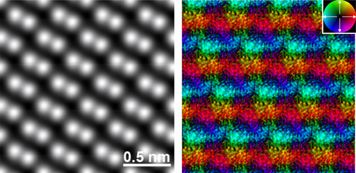
Figure 1. Real-space magnetic field image of an antiferromagnetic α-Fe2O3 The atomic structure image (left) and corresponding magnetic field image (right). In the atomic structure image, Fe atoms are visualized as bright spots. In the magnetic field image, the color contrast indicates the magnetic field orientation and strength. The inset color wheel indicates how color and shade denote the magnetic field orientation and strength in the vector color map. The antiparallel magnetic fields on the adjacent Fe atomic layers are clearly observed, visualizing antiferromagnetic order in this crystal. © Naoya Shibata
-
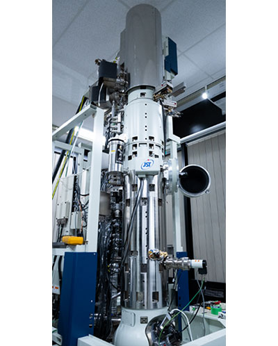
Figure 2. Developed Magnetic-field-free Atomic-Resolution STEM (MARS) A newly developed magnetic object lens system is installed in this microscope. Combined with a higher-order aberration corrector, an electron beam can be focused on a sample at atomic scale, while keeping the sample in a magnetic-field-free condition. © Naoya Shibata
Program Information
- JST SENTAN
- Research Area “Development of Advance Measurement and Analysis Systems”
- Research Theme “Development of atomic-resolution, magnetic field-free electron microscope”
- JST PRESTO
- Research Area “Precise Arrangement of Atoms and Molecules and Its Properties and Functions”
- Research Theme “Development of Ultra-Low Electron Dose STEM Technique and Structural Analysis of Atomic and Molecular Arrangements in Real Space”
Journal Information
Yuji Kohno, Takehito Seki, Scott David Findlay, Yuichi Ikuhara and Naoya Shibata. “Real-space visualization of intrinsic magnetic fields of an antiferromagnet”, Nature. Published online February 9, 2022, doi:10.1038/s41586-021-04254-z
Contact
-
[About Research]
Naoya Shibata
Professor, Institute of Engineering Innovation, School of Engineering,
The University of Tokyo
E-mail: shibata sigma.t.u-tokyo.ac.jp
Takehito Seki
Assistant Professor, Institute of Engineering Innovation, School of Engineering,
The University of Tokyo
E-mail: sekisigma.t.u-tokyo.ac.jp -
[About Program]
Junichi Hoshi
Department of Industrial-Academia Collaboration, JST
E-mail: sentanjst.go.jp