Progress Report
Challenge toward the Control of Intractable Cancer through Understanding of Molecular, Cellular, and Interorgan Networks[2] Technology development for integrated analysis and verification of patient biometric data
Progress until FY2024
1. Outline of the project
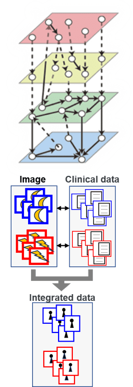
By utilizing the patient biospecimen bank, in addition to parent-derived genomic data, various data such as gene mutation and gene expression at the lesion site can be obtained. However, the utilization technology is currently very underdeveloped. In this theme, we will develop an integrated analysis method to reveal the key molecules and networks (molecules and cell/tissue/organ networks) in the onset process from "multi-layered data" derived from patients.
Since the amount of patient samples is very small, there is a big limitation as an experimental material. In order to overcome this, we will further evolve animal models and patient organoid models. By using patient organoids, it is possible for the first time to analyze biological responses to drugs and gene mutations. It also has great potential as an optimal drug selection system for individuals.
In addition to acquiring sequential data over time, imaging technology has the potential to lead to non-invasive diagnosis in the future. In this theme, we will proceed with the development of imaging technology (sensors and probes).

2. Outcome so far
Construction of an integrated analysis platform for "multi-layered data"
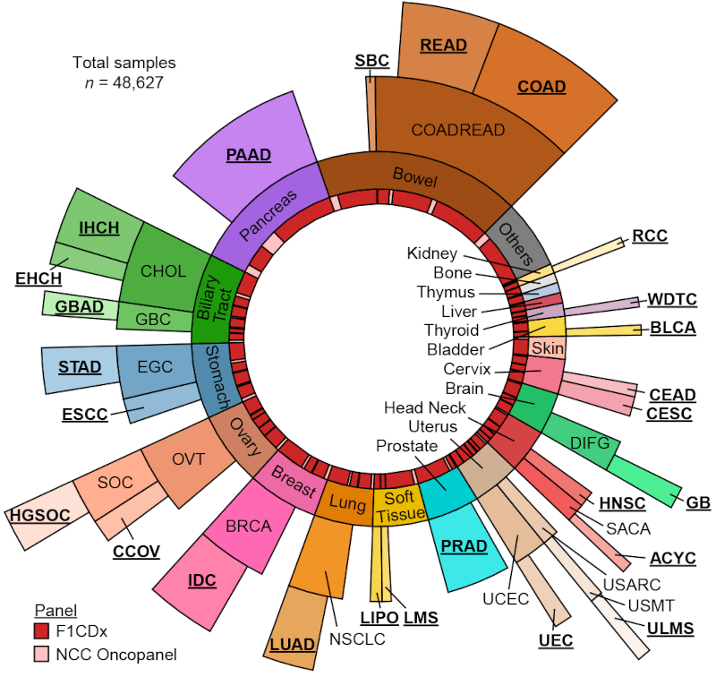
Using mouse omics data, we have developed a technique for estimating multi-hierarchical networks. We have also investigated pseudo-time analysis that arranges patient organoids at various stages along the path of onset. We have developed an exploratory image analysis technique using deep learning to predict the prognosis of pancreatic cancer from peritoneal washing cytology images. Furthermore, we have established a method for analyzing genomic abnormalities in Japanese cancers across cancer types.


Development of next-generation organoid system
We proceeded with the establishing of new mouse models with pancreatic cancer. We have also developed a next-generation organoid culture method using iPS cells and elucidated the prediction of progression risk due to single nucleotide polymorphisms in glucose metabolism genes. Furthermore, we have succeeded in generating liver organoids with functional multi-layered structures. This will be a new foundation for disease research, drug evaluation, and regenerative medicine.

Building an imaging analysis platform
We have built an imaging analysis platform that researchers can use collaboratively.
Retrospective analysis of CT images revealed that focal pancreatic atrophy can be observed several years before the onset of pancreatic cancer.
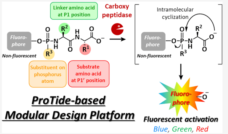
Using a modular design platform, we have developed a fluorescent probe for proteolytic enzymes, which makes it possible to detect enzyme activity in cancer tissues by fluorescence. This is expected to be applied to new diagnostic drugs to find cancer sites during surgery.


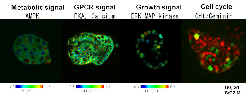
Live imaging analysis of pancreatic cancer patient organoids expressing biosensors revealed heterogeneous dynamics of ERK and AMPK activity.
3. Future plans
Utilizing the established organoids from very early and advanced cancer and mouse models, we will proceed with the development of integrated analysis technology for "multi-layered data." We will promote the development of next-generation organoid technology specialized for cancer research. We will continue our efforts to advance imaging technology. We will also start probe screening using clinical samples.