Research Results
The world's first success in photographing the division of a fertilized egg of a plant!
Real-time Observation of Plant Cells FY2017
- Tetsuya Higashiyama (Professor, WPI Institute of Transformative Bio-Molecules, Nagoya University)
- PRESTO
- The Dynamic Mechanism of and Fundamental Technology for Biological System "Dynamic System of Pollen Tube Guidance" Researcher (2007-2011)
- ERATO
- "HIGASHIYAMA Live-Holonics Project" Research Director (2010-2016)
The live imaging technology to observe the cell division process of a plant fertilized egg
A fertilized egg of an animal can be easily removed, and the process of cell division can be observed alive. However, a fertilized egg of a plant cannot be removed and observed alive since it lies deep inside of a pistil. No one has ever seen the fertilization process or the cell division process of an angiosperm, a flowering plant.
Professor Tetsuya Higashiyama developed a system that enables the real-time observation (live imaging) of a living fertilized egg through the ERATO project, and succeeded in taking the world's first live images that clearly show the fertilized egg of a plant dividing to form an embryo. The motto of this project is to establish the"live cell (living cell) biology", in which living cells and molecules of multicellular organisms can be flexibly manipulated and analyzed under a microscope Professor Tetsuya Higashiyama stated that it was his very big goal to observe and explore interactions between components and the whole of a living body with the live cell technology.
Live imaging of fertilized egg division and embryogenesis of Arabidopsis thaliana
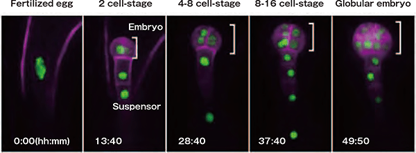
A fertilized egg divides to form an embryo and a suspensor. The sections indicated in green represent the cell nucleus and those in pink represent the cell membrane. Embryonic cells divide to form globular tissue changing the direction of division, while suspensor cells divide along the longitudinal direction only to form a rod-shaped tissue.
Laser microscope and micro devices that support the study
The two main pillars, "laser microscope" and "micro devices", played an important role in the study. Professor Higashiyama organized a project team consisting of experts in various fields including optical equipment, engineering, and information science.
A laser microscope uses a laser beam with a shorter wavelength and higher rectilinearity than the visible light of an optical microscope, allowing the acquisition of sharper and more high-contrast images. This laser beam also enables cell manipulation; for example, it can destroy target cells or parts of cells, allowing the investigation of cell functions. The other main pillar is the application of new micro devices produced by micromachining technologies. Micro devices, tiny devices called"tip,"are produced by engineering technologies, and have made it possible to grow plant cells in a form that is appropriate for effective culture/observation. The latest micropillar arrays (a structure in which many tiny pillars are arranged at uniform distances) have a flexible structure, which enables the observation of delicate changes in the cells.
The discovery of the "cell regenerative ability" and "new cell fusion phenomenon" of plants
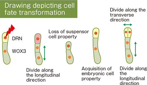
One of the remarkable achievements in this project is the elucidation of the"regenerative ability of cells."A fertilized egg of an angiosperm divides asymmetrically into apical cells (eventually developing into the plant body) and basal cells (responsible for supplying nutrients to the embryo). The research group destroyed apical cells with the laser technology described above, and observed the subsequent influence on the basal cells sequentially. As a result, surprising facts that had not been known to date were revealed. When apical cells were damaged, "cell fate transformation" occurred where basal cells, which had already been developing into suspensor cells, played the role of apical cells to compensate for the damaged apical cells. This is a proof of the surprising regenerative ability of plants.
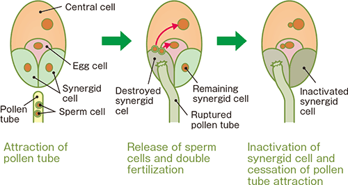
Furthermore, a completely new type of cell fusion phenomenon was discovered. In the development process of a seed of a plant, a pollen tube that extended from pollen is attracted by a tissue from which a seed is developed. At this time, what attract the pollen tube into the tissue are two synergid cells that lie adjacent to the egg cell. After the pollen tube reaches one of the synergid cells, two sperm cells gush out of the tip of the pollen tube, destroying the synergid cell. Then, one of the sperm cells is fertilized with the egg cell to develop into the embryo, while the other is fertilized with the central cell to develop into endosperm. Professor Higashiyama and his colleagues studied the changes that occurred in the remaining synergid cell by using Arabidopsis thaliana, a major model plant. As a result, a cell fusion phenomenon was observed in which the synergid cell and endosperm were fused and their contents were mixed. This phenomenon caused the rapid dispersion of the attractants in the synergid cell, suppressing the attraction of a pollen tube. This study elucidated the cessation mechanism of pollen tube attraction.
The cell fusion of plants has not been known to date except for fertilization, so this discovery not only greatly changed the perspective on plant cells, but also presented a new function of plant cells. It is no exaggeration to say that this discovery will break out the paradigm.
Changes in cells caused by the destruction of an apical cell
Double fertilization and cell fusion of synergid cell in an angiosperm
Contribution to the accelerated progresses of global embryogenesis study
The progresses of the global study of embryogenesis will accelerate as a result of the live imaging technology of plants. It is expected that the elucidation of the mechanism that enables cell fate transformation will contribute to the development of breeding/culture technologies associated with plants, including efficient tissue culture. The presence of the program that causes cell fusion other than fertilization was shown in plants, which will lead to the development of new cell fusion technologies of plants. In addition, Professor Higashiyama and his colleagues succeeded in developing the reagent "ClearSee," which makes a plant transparent, allowing detailed observation of the inner structure of a plant.
The series of studies described above greatly shocked the whole world. Due to the close cooperation among experts in imaging, engineering, and information science fields, the study is progressing further toward the identification of key molecules. Further achievements are expected in the future.