Under JST’s Strategic Basic Research Programs, Noda Nobuo (Laboratory Head) and Fujioka Yuko (Senior Researcher) of the Institute of Microbial Chemistry, in collaboration with other researchers, discovered that a liquid-like condensate (liquid droplets1)) in which the Atg protein is clustered through the liquid-liquid phase separation2) is the structure responsible for the progression of autophagy.
Autophagy is one of the mechanisms through which cellular protein is degraded. Previously, it was known that Atg proteins assemble to form a structure called PAS3). However, the mechanism through which Atg proteins assemble and the physicochemical property of the formed structures had been unclear.
The research team elucidated characteristics of PAS through observing the Atg protein using a fluorescence microscope and successfully reconstituted PAS in vitro. The team revealed, for the first time, that PAS is in the state of liquid droplets formed by liquid-liquid phase separation of Atg13 together with other Atg proteins and that this liquid droplet is responsible for autophagy.
The finding that liquid-liquid phase separation directly controls autophagy suggests its involvement in a wide range of intracellular life phenomena. Reconsideration of molecular mechanisms underlying various intracellular phenomena is expected to proceed. Moreover, development of autophagy-specific control agents that focus on the regulation of liquid-liquid phase separation in autophagy-related diseases is anticipated.
1) Liquid droplet
A condensate of macromolecules with fluidity created when protein and/or nucleic acids undergo liquid-liquid phase separation. The droplets are also known as “membraneless organelles” and perform various functions within the cell. A droplet spontaneously assumes a spherical form. It also has high internal fluidity, and it actively exchanges molecules with its surroundings.
2) Liquid-liquid phase separation
This is the phenomenon of a uniform liquid phase separating into multiple liquid phases. It is observed in daily life as the separation of water and oil, and occurs within cells with proteins and nucleic acids.
3) PAS (Pre-autophagosomal structure)
The collected structure that the Atg protein forms near the vacuole under nutrient starvation in yeast is called PAS. It is assumed that autophagosomes formation starts at PAS.
-
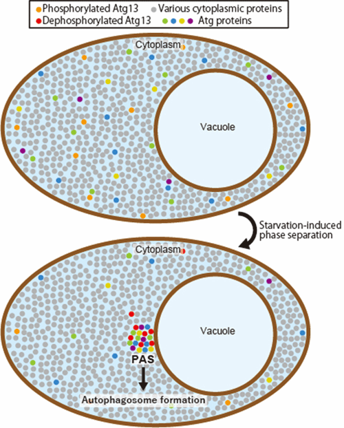
Figure. Model of PAS organization via liquid-liquid phase separation Under nutrient-rich conditions, Atg proteins are dispersed and mixed with various proteins in the cytoplasm. Upon starvation, Atg13 is dephosphorylated, which triggers the phase separation of Atg13 with other Atg proteins to form a liquid droplet on the vacuolar membrane, where autophagosome formation proceeds.
Program Information
- JST CREST
- Research Area “Structural Life Science and Advanced Core Technologies for Innovative Life Science Research”
- Research Theme “Structural life science for unveiling membrane dynamics during autophagy”
Journal Information
Yuko Fujioka, Jahangir Md. Alam, Daisuke Noshiro, Kazunari Mouri, Toshio Ando, Yasushi Okada, Alexander I. May, Roland L. Knorr, Kuninori Suzuki, Yoshinori Ohsumi and Nobuo N. Noda, Nature. Published online February 5, 2020, DOI: 10.1038/s41586-020-1977-6
Contact
-
[About Research]
Noda Nobuo
Laboratory of Structural Biology, Institute of Microbial Chemistry
TEL: +81-3-3441-4173
E-mail: nn bikaken.or.jp
[About Program]
Kawaguchi Tetsu
Department of Strategic Basic Research, JST
E-mail: crestjst.go.jp