Progress Report
Development of a Neuroscientific Basis for Visualization and Manipulation of the Mind[2] Optical manipulation of functional brain networks by optogenetics
Progress until FY2024
1. Outline of the project
This research aims to understand how mental states—what we call the "mind"—are connected to the brain by using a technique called optogenetics, which allows scientists to control brain activity with light. Using advanced holographic microscopes, we shine light on specific networks in the brain of a mouse while it behaves naturally. By observing how the mouse’s behavior or mood changes in response, we are beginning to uncover how brain activity shapes the mind.
2. Outcome so far
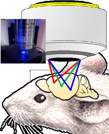
We developed a system that can stimulate multiple parts of the mouse brain at once, even from outside the skull. This is done using a 3D holographic light stimulation system (right upper figure) which delivers light with pinpoint accuracy to many neurons simultaneously—up to 1,350 cells at once.
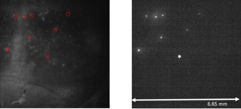
To make the light reach only the right areas, we applied fluorescent beads to the mouse’s skull and used the way light scatters to fine-tune the focus (right figure). This technique, called phase-conjugate illumination, enables us to target specific cells even through the skull. Experiments with real mice confirmed its effectiveness.
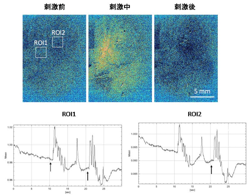
We also built an optical system that uses two different wavelengths of light—one for observing brain activity (red) and one for stimulating it (blue)—without interference. To run these tests, we created a special type of mouse by combining four genetically engineered lines. This “quadruple transgenic mouse” allows both observation and stimulation of brain networks in the same animal. Using this system, we discovered that certain inner brain areas—such as the prefrontal cortex and cingulate cortex—can trigger voluntary walking when stimulated. When this behavior occurred, brain activity across the entire cortex became highly synchronized. However, when no behavior change was seen, brain network changes remained minimal. This shows that the brain’s response to external stimulation can vary greatly.
3. Future plans
In the next stage of our research, we will use this system to precisely control brain networks in the cerebral cortex and examine how those manipulations affect behavior and internal mental states. By doing so, we aim to pinpoint which brain networks are responsible for changes in emotion and behavior. Ultimately, this research could provide new insights into how the brain gives rise to the mind and help lay the groundwork for future treatments of mental and neurological disorders.
(MATOBA Osamu, TAKUMI Toru: Kobe University)