Research Results
Preventing the release of gas or water even in a high vacuum
Observing organisms in a living state with nano-suits FY2016

- Takahiko Hariyama (Professor, Department of biology, Hamamatsu University School of Medicine)
- CREST
- Establishment of innovative manufacturing technology based on nanoscience
"Novel Engineering of Hierarchically Structured Biomimetic Surfaces" Co-researcher (2008-2014)
A shift from the observation of"deformed and non-living organisms"to the observation of"actual living organisms"
In order to observe the micro structure of the surface of an organism, it is necessary to arrange the sample in the high vacuum chamber of an electron microscope so that an electron beam can be sent to the sample easily. If you do so, however, what will happen to an organism of which water accounts for 80% of its body weight? In order to adjust to various environments, the body surface of an organism is covered with an extracellular substance (ECS: a substance formed by the accumulation of substances secreted from a cell); however, under an extreme situation like a high-vacuum state, the ECS is not able to prevent the release of gas or water. Understandably, the external structure of the organism will become significantly deformed due to volumetric shrinkage caused by dehydration, resulting in the death of the organism. Therefore, it's still the case that when observing the micro structure of the surface of an organism with an electron microscope, researchers apply a drying treatment and surface metal coating to a "lifeless organism"after chemically fixing it, in order to keep the state of the organism as close to the living state as possible.
In April 2013, however, it became possible to observe a "living organism" with an electron microscope. The group led by Professor Takahiko Hariyama, a co-researcher of a project adopted in CREST in 2008 (chief researcher: Professor Masatsugu Shimomura at Chitose Institute of Science and Technology) succeeded in developing a "nano-suit" (nano polymer membrane) that prevents the release of gas and liquid from an organism even in a high vacuum, thus protecting its life by irradiating a plasma or electron beam to the ECS or a thin liquid membrane mimicking it.
A thin membrane was formed by means of an electron beam or plasma irradiation
First, the research team observed various organisms using a high-resolution scanning electron microscope (FESEM) and found that most of them died and their surface structure had become greatly deformed. However, some organisms (including drosophila larvae) that have an ECS with a high viscosity in the outermost layer remained active in the microscope (larvae A, B and C) without suffering neither shrinkage nor deformation of their micro structure. On the other hand, however, the larvae that were observed after being left to stand for one hour without receiving electron beam irradiation were found to have been squashed and dead due to dehydration (larvae E, F and G).
With that, the researchers observed the ultrathin cut section of the outermost layer of larva C with a transmission electron microscope, and found that a thin membrane with the thickness of 50 ‒ 100 nanometers (nm: a nm is equal to one billionth of a meter) was formed (D). In contrast, a thin membrane was not found in the case of larva G (H). As a result, it was found that a thin membrane was formed on the outermost layer of the larvae by means of electron beam irradiation and this membrane was inhibiting the release of gas and liquid. The same result was also obtained in the experiment using plasma irradiation. Thus, it was confirmed that "an ECS on the outermost layer of a larva forms a thin membrane with the thickness of 50 ‒ 100 nanometers that can inhibit the release of internal substances under the electron beam or plasma irradiation, which brings the FE-SEM observation under high vacuum to realization." The research team named this thin membrane"nano-suit"(polymer membrane of nano-scale).
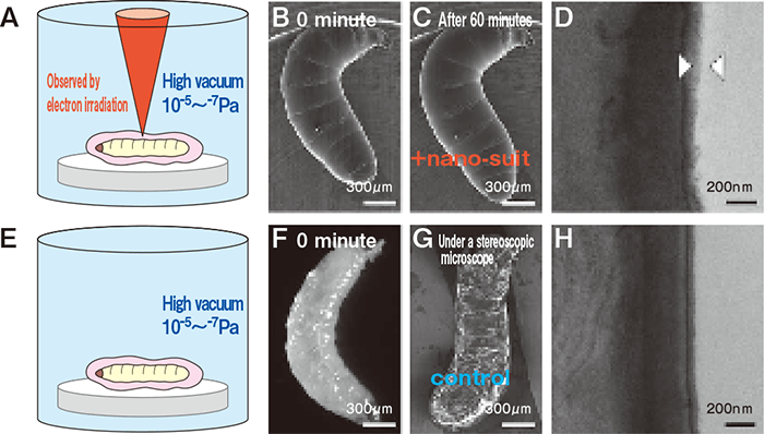
Drosophila larvae were directly put in the electron microscope and observed
Developing a nano-suit by irradiating a surfactant with plasma
The next step was the development of a "nano-suit." After analyzing the components in an ECS, the research team selected a solvent having a chemical functional group similar to that of an ECS and conducted a test on organisms that don't have an ECS to see if they would express the similar functions. What was selected was a surfactant "Tween 20" which is designated as a food additive. They applied this to mosquito larvae in a thin layer and then irradiated them with plasma for a few minutes (In a normal FE-SEM observation, mosquito larvae get squashed and die within a few minutes. larva A). Through the FE-SEM observation, it was found that the samples with a nano-suit were, however, staying active under a high vacuum without experiencing any morphological change (larva B). A thin membrane with the thickness of 50 ‒ 100 nm was formed on the outermost layer of the larva (wiggler) after the observation as in the case of the ECS of drosophila larvae. The nano- suit created with Tween 20 was proved to be able to fulfill its function and bring the observation of microstructure of organisms in a living state to reality. The nano-suit method was developed in this way; subsequently, this method was proven be able to be performed with surfactants other than Tween 20.
The nano-suit method was applied to various organisms that are small enough to be put under an electron microscope, most of which could sustain their lives and keep active.
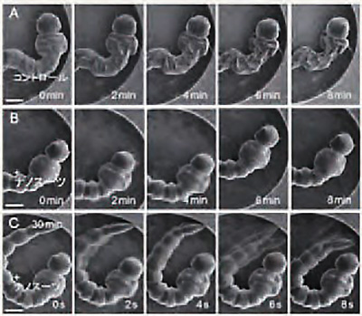
You can observe organisms covered with a nano-suit moving actively without experiencing a morphological change. C stayed active for more than 30 minutes.
From microscopic "biomimetics" to the immense world of "life science"
The"nano-suit method"developed by the research group led by Professor Hariyama has made it possible to observe the microstructure of organisms in a living state, which is sure to contribute to the advancement of research fields including the fields of "monozukuri (manufacturing)," and especially the field of "biomimetics." This is a technological field in which various studies are conducted with the aim of creating new materials or systems through learning and mimicking the functions and mechanisms that organisms have developed to survive in various environments. Increasing attention is paid to the development of materials that mimic the microstructure of organisms.
Some well-known areas might be studies on the super water repellency of lotus leaves, the structural color of butterfly wings and the low frictional properties of shark skin.
Meanwhile, the nano-suit method has made it possible to directly observe the activities being done in extremely microscopic areas in small animals, cells, etc. It is also expected that such observation will lead to understanding phenomena or behaviors relating to organisms that are not currently understood, in addition to revealing the interaction between tissues and cells. Thus, the creation of the nano-suit method will eventually lead to a contribution to the development of immense"life science"that includes biology, agriculture and medicine, etc.
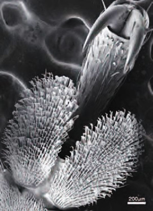
High resolution photographs taken under a high vacuum state of the microstructure of the front leg of a leaf beetle to which nano- suit method was applied