Research Results
Combining the optical microscope and the mass spectrometer
Imaging Mass Microscope FY2016
- Mitsutoshi Setou (Professor, Department of Cell biology and Anatomy, Hamamatsu University School of Medicine)
- Development of Advanced Measurement and Analysis Systems
- System Development "Development of mass microscope" Team leader (2004-2008)
- Kiyoshi Ogawa (General manager of advanced technology development, Technology Research Laboratory, Shimadzu Corporation)
- Development of Advanced Measurement and Analysis Systems
- Practical Realization "Development and application of mass microscope" Team leader (2009-2011)
An innovative discovery about the pathological change of'abdominal aortic aneurysm'
There is a disease called'abdominal aortic aneurysm.' When someone suffers from this disease, part of the vascular wall becomes swollen and bulges in a part of their aorta that runs through their abdomen. Such a bulge is fragile and it can easily rupture. What is more, once it ruptures, it results in death from excessive bleeding in 80 percent of the patients. However, since the cause and the mechanism of the bulge formation is unknown, treatments are limited to surgical ones, such as removing lesions. No effective medical prevention or treatment method is known at this time.
In September 2012, a discovery that could lead to the breakthrough in learning how the formation of the abdominal aortic aneurysm occurs was reported from the research group of Professor Mitsutoshi Setou et al. As a result of analyzing a lesion section that had been removed through surgery, a pathological change was confirmed; Blood vessels inside an aortic wall that formed a bulge became narrow and thus the blood volume decreased. This indicates the possibility that vascular walls can become fragile when oxygen or nutrition does not reach them due to a decrease in blood flow inside the vascular walls.
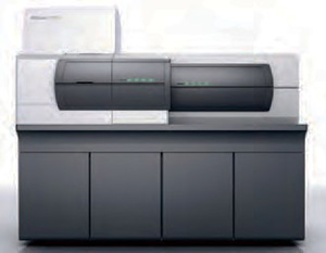
It is the'imaging mass microscope,'that helped derive this discovery. This microscope was created by combining an optical microscope and a mass spectrometer. This is a new measuring and analyzing apparatus developed by the group of Professor Setou and the project team of Mr. Kiyoshi Ogawa (General manager of advanced technology development) at the Shimadzu Corporation as part of the development of advanced measurement and analysis systems In April 2013, this imaging mass microscope was produced (and introduced) with the name 'iMScope.'
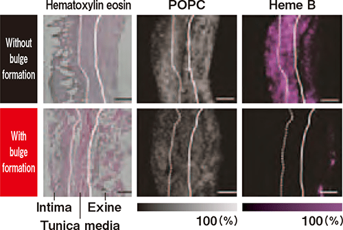
(Left column) Pathological images by a pre-existing microscope
(Center and right column) Imaging pictures by mass microscope
As for the pathological images and POPC (distribution of lipid) images, no big difference was observed regardless of whether there was the existence of a bulge or not. On the other hand, a large amount of heme B (molecules that signify blood) existed where there was no bulge, whereas, it was rarely seen around the abdominal aorta wall where a bulge was formed.
Combination of the'observation'of pathological tissues and the'mass spectrometry'of molecules
According to Professor Setou, who has taken the lead in the development of mass microscopes since 2004 as a team leader of JST Development of Systems and Technologies for Advanced Measurement and Analysis, this development was based on a new concept of 'molecular biology' that was introduced in the medicine field in the 1990s. This biology explains life phenomenon from the viewpoint of molecules.
The world of medicine has been developing through systematically constructing enormous amounts of knowledge that is empirically gained, as well as with pathology that'acknowledges patterns of lesion sections' using microscopes. In addition to these, the viewpoint that'a human body is made from molecules' has been accepted. In other words, a new method of'specifying what kind of molecules make up a particular kind of tissue'is added to the pre-existing method of 'observing pathological tissues.' In order to establish 'what kind of molecules make up a particular kind of tissue' or elucidate various types and properties of molecules included in samples, it is appropriate to use 'mass spectrometry,'which elicits the answer by measuring and analyzing the mass.
By combining this pathological 'observation' and 'mass spectrometry' that brings understanding from the viewpoint of molecular biology, this concept was born: Visualization like a map pertaining to the existence of molecules with the imaging mass microscope.
Analyzing molecules by ionization, and detecting distribution with images
In order to measure targeted molecules with the pre- existing mass spectrometry method, pre-treatments are needed; Samples should be ground down and isolated per component before analysis. If whole tissue is ground down for analysis, it is then impossible to know where the tissue originated from. That is to say, even if specific molecules accumulate at high-density on a specific section of a sample, we lose such information when we grind it down. It was difficult for researchers to perform 'image observation'and'mass spectrometry' at the same time; They could not observe samples by leaving the molecules where they were originally located. The researchers must have been frustrated by this dilemma.
However, if a researcher uses a mass microscope using laser, it is capable of irradiating laser beams directly on an observation sample for ionizing and imaging molecules in order to analyze them. The development project adopts Matrix Assisted Laser Desorption/Ionization (MALDI) (the method which enhanced the technique developed by Mr. Koichi Tanaka at Shimadzu Corporation who won the Nobel Prize for Chemistry in 2002), which can analyze by ionizing plural molecules with laser irradiation directly on the sample, and can even compare their distribution with one another using images. This can be called a 'molecule map.'
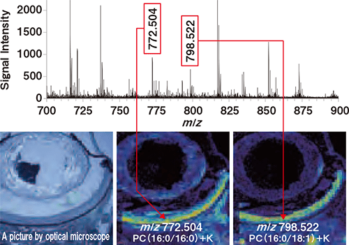
Right-side two pictures show image data that indicates distribution of molecules with specific mass, or 'molecule maps.' Left-side picture is an image obtained by an optical microscope.
Increasing expectation as an analysis tool in various fields
The new measurement analysis apparatus 'iMScope' which is equipped with a high-performance optical microscope and provides pictures obtained by imaging mass analysis under atmospheric pressure has been contributing to researches especially in the field of basic medical sciences. When it comes to the 'abdominal aortic aneurysm, the researchers discovered that blood volume had decreased where the lesion had formed. This result indicated the possibility below: Control advanced / relapsed aneurysm by improving the bloodstream. Prevent aneurysms with medicines that lessen inflammation and prevent blood vessels from being clogged. Based on this knowledge, Hamamatsu University school of medicine has launched a clinical research for the treatment and prevention of the abdominal aortic aneurysm. Furthermore, this knowledge is now being utilized for the research and development of medical supplies, such as verification of the process of medicine metabolism and accumulation. The expectation as an innovative analysis tool is increasing in other fields like food safety, quality control of organic materials, and etc.
Imaging mass microscope 'iMScope'