Research Results
Whole-body imaging at a single cell level!
A whole mouse made transparent FY2016
- Hiroki Ueda (Professor at the Graduate School of Medicine, The University of Tokyo / Group Director at Quantitative Biology Center, RIKEN)
- CREST
- Innovation for Ideal Medical Treatment Based on the Understanding of Maintenance, Change and Breakdown Mechanisms of Homeostasis among Interacting Organ Systems
"Challenge to Reveal Dynamic Properties in Circadian Sleep-Wake Homeostasis" Research Director(2013–2018)
Challenge towards the dream of seeing every single cell in the body
Although about 350 years have passed since Robert Hooke of England discovered cells under a microscope, no-one in the world has seen and identified every single cell in the bodies of humans or mice as a high resolution image. The body of a mouse with body weight of 30 g is made up of about 30 billion cells, and complex cellular network spreads across the entire body. Even if we focus on brain as a critical organ, a brain with 0.5 g of weight contains nearly 500 million cells. Observing the individual cells is more difficult than observing individual persons from outside the islands of Japan. What is then needed to observe the individual cells in further detail? Collaborative research group led by Professor Hiroki Ueda has tackled this problem as a part of CREST "Innovation for Ideal Medical Treatment Based on the Understanding of Maintenance, Change and Breakdown Mechanisms of Homeostasis among Interacting Organ Systems" project. In order to comprehensively analyze the entire body as a single system, they have attempted to develop an imaging technology to obtain the function of the cell network and genes across the body as a 3D image with single-cell level resolution.
Figure 1
Entire mouse made transparent (the left figure shows the infant mouse, the right figure shows the adult mouse)
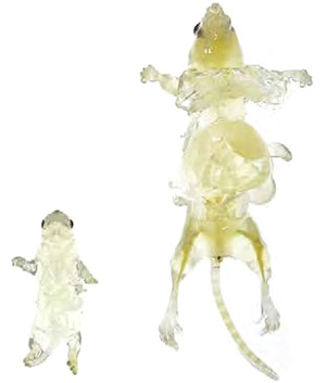
Image provide by Riken
Removal of endogenous pigments as requirement for transparency
In the observation of brain cells, the light is scattered by water, protein, and lipid in the cells, making it unclear to see. Professor Ueda thought that" if it cannot be seen, it should be made see-through." He has removed the lipid which causes the scattering of light and made the refractive index in the tissue uniform, and succeeded in obtaining a highly transparent brain sample.
However, an effective method was needed for the removal of endogenous pigments that absorb lights in order to achieve transparency in organs with high content of endogenous pigments such as a heme in red blood cells, for example liver and spleen. Many researchers have attempted to make the organs transparent in the past; however no efficient method was discovered for the removal of pigment in tissues containing endogenous pigments without disruption of proteins.
At last, the organs and the body is made entirely see-through!
The solution to this problem was the transparency reagent (ScaleCUBIC reagent, hereinafter referred to as CUBIC reagent) used in CUBIC*, which Professor Ueda and his colleagues developed as whole-brain imaging and analyzing technology. This reagent was accidently discovered to efficiently decolorize blood. They have clarified the mechanism where aminoalcohol, a component in CUBIC reagent, efficiently dissolves the red pigment heme in the blood, resulting in decolorization of the blood.
Furthermore, a new perfusion protocol using CUBIC reagent was developed in order to obtain a sample with higher transparency. The procedures involved are as follows: Firstly, perfusion fixation is performed on the body of a mouse using 4% paraformaldehyde (typical method of fixation to maintain the components in the tissues in a state as close to the living state as possible). Following this, CUBIC reagent diluted to 1/2 concentration is circulated to the whole body via the vascular system. The heart, lung, kidneys, liver, pancreas, spleen, muscle, stomach, intestinal tract, and skin are immersed in the same reagent for 10 days. This process enables the entire organ to become transparent. If the body of a mouse with the skin removed is immersed in the reagent for 2 weeks, the whole body can be made transparent.
Using these transparent organ and body samples, and by using the light sheet fluorescence microscopy (microscope where laser ray is spread in a sheet-like manner and radiated from the side of the sample and the image is taken from the top to enable a certain plane in a sample in a single session (refer to Figure 2)), it is possible to obtain a single-cell resolution 3D imaging data of an entire organ or body, showing the positions of the cells, etc., in a short period of 1 hour. In previous methods, high resolution imaging of an entire brain required at least a day. Using this microscope where the radiating axis and observing axis cross orthogonally, a 2D image can be obtained quickly, and a 3D image can be obtained at high-speed by simply changing the sheet height.
Adaptation of high-speed 3D imaging in Figure 2
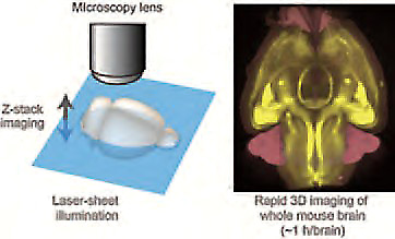
Image provided by Riken
Use in biology and medicine, as well as elucidation of sleep-wake cycles
This technology by Professor Ueda and his colleagues can be applied to 3D pathological analysis and 3D anatomy, as well as immunohistological analysis. Since this technology enables the clarification of vital phenomenon and its operating principles on an individual body level, it is expected to contribute not only in biology but also in the field of medicine. This technology also plays an important role in Professor Ueda's ongoing research on the sleep-wake cycles. It will help investigate the activity of individual cells important in the research, thereby assisting in clarifying the mechanisms involved. It is said to eventually elucidate and treat the psychiatric disorders of depression and schizophrenia. The collaborative research continues its challenges for dreams further ahead.
* Abbreviation of"Clear, Unobstructed Brain/Body Imaging Cocktails and Computational analysis."It is a combination of clearing reagents and protocols for whole-brain imaging, and computer image analysis.
Figure 3 3D anatomy using CUBIC
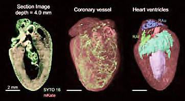
The characteristic structures of the organ can be extracted by image analysis. In the heart, internal structures such as ventricles and atria were extracted. Image provide by Riken