Research Results
Successful measurement leading to improvement of the accuracy of epilepsy diagnosis
Paving the way for practical application of the “magnetoencephalography system that can be used anywhere,” allowing downsizing and mass production of the deviceFY2024

- Development and implementation company: Konica Minolta, Inc.
Research leader: ANDO Yasuo (Professor, Tohoku University) - Strategic Promotion of Innovative Research and Development (S-Innovation) Program
- “Development of a simultaneous room-temperature-measurement system of magneto-cardiography, magneto-encephalography and magnetic resonance imaging with tunnel magneto-resistance devices” (2011-2020)
The world's first successful measurement of very weak magnetoencephalography (MEG) signals
The magnetoencephalography system, which measures the magnetic field generated by weak electrical currents that flow in the neurons of the brain (magnetoencephalography: MEG), has an important role in the diagnosis, etc. of epilepsy*1. The group of Prof. Nobukazu Nakasato (Department of Epileptology, Graduate School of Medicine, Tohoku University) and Prof. Yasuo Ando (Department of Applied Physics, Graduate School of Engineering, Tohoku University) was the first in the world to successfully measure very weak MEG signals using contact-type sensors that utilize the tunnel magneto-resistive (TMR) effect*2.
This enabled the creation of a measurement device with high accuracy that is much more compact than devices with conventional superconducting sensors and is operable at room temperature. Thanks to low manufacturing costs, mass production is possible. Therefore, it is expected to pave the way for future development of a portable “MEG system that can be used anywhere,” which can easily be used in any environment.
*1 Epilepsy
“Epileptic seizure” refers to a condition caused by excessive excitation of cerebral neurons that occurs
due to various causes, resulting in convulsions of the limbs and face, etc., sensory symptoms, and
autonomic symptoms.
*2 Tunnel magneto-resistive (TMR) effect
This refers to a phenomenon consisting of changes in the electrical resistance of two ferromagnetic layers
that lie on either side of a thin insulating layer induced at room temperature by their respective
directions of magnetization. It was discovered in 1995 by Terunobu Miyazaki (Emeritus Professor, Graduate
School of Engineering, Tohoku University).
Device-related problems that delayed the spread of MEG systems
While seizures can be controlled by continuous medication in about 70% of epileptic patients, surgical resection of the causal site of the brain is effective for refractory epilepsy, which does not respond to medication. The MEG system is a device that measures magnetoencephalography, which can be defined as “magnetically measured brain waves.” Its excellent property is its higher measurement accuracy compared to electrical measurement of brain waves, allowing measurement of the active sites in the brain in millimeter units. Thus, MEG is very effective for determining the site to be resected in surgical treatment of epilepsy. In addition, it has been utilized for diagnosing various brain functions.
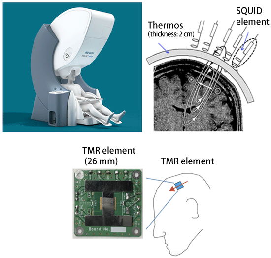
However, the superconducting sensors used in conventional MEG systems needed to be cooled with ultracold liquid helium to ensure full performance of the sensors, inevitably resulting in upsizing the device. In addition, it was necessary to insert the head into a helmet-type device with a fixed shape adapted to a thick helium container, which prevented full accuracy from being obtained because the magnetic field sensors could not be placed close enough to the scalp (Fig.1). These superconducting sensors are susceptible to environmental magnetic noises, and consequently can be used for measurement only in a special magnetic shielding room that blocks external magnetism. Moreover, liquid helium is expensive, and its cost alone represents tens of millions of yen annually. Thus, these various device-related problems were hindering the spread of the MEG system.
Prof. Nakasato is a pioneer who has been engaged in the research of magnetoencephalography at Tohoku University since 1987. When he was searching for a way to solve the device-related problems mentioned above, he encountered the research of TMR elements, which the group of Prof. Ando and collaborators had been developing jointly with Spin Sensing Factory and Konica Minolta. This encounter led to the research outcome reported here, namely, the development of an epoch-making MEG system.

Fig.1 Comparison between old and new MEG systems
Measurement of the magnetic field using conventional superconducting sensors (upper) requires a thermos structure with a thickness of 2 cm for cooling with helium, which hindered high-accuracy measurement because it could not adhere to the patient’s head. By contrast, the magnetic field measurement (lower) based on the TMR element used in this research operates at room temperature and can be placed in contact with the scalp to perform recording.
Research results leading to practical application of the TMR-MEG system
This research was conducted using spintronics technology, which is Tohoku University’s area of expertise. This is a new engineering research field, which combines electrical engineering (electronics) and magnetic engineering (magnetics). The TMR element was produced using spintronics technology. The electron also has a property of micromagnet, called “spin.” Spintronics is a technology utilizing the spin property.
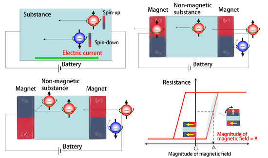
The TMR effect, which led to the present development of the contact-type sensors of the MEG system, refers to a phenomenon consisting of changes in the electrical resistance of two ferromagnetic layers that lie on either side of a thin insulating layer induced at room temperature by their respective directions of magnetization (Fig.2). When the magnetization of one of the ferromagnetic layers is changed by the external magnetic field, this can be measured as weak electrical resistance, which led to development of a high accuracy magnetic sensor with very low power consumption. The TMR magnetic sensor utilizes this.
Using TMR elements, the research group was the first in the world to successfully measure very weak MEG signals generated by the somatosensory cortex of the right cerebral hemisphere when the median nerve*3 in the left wrist of a healthy subject was electrically stimulated (Fig.3). Clinical application of conventional MEG systems is most advanced in an examination method consisting of measurement of MEG signals elicited when sensation on the skin and mucosa reaches the cerebrum to be “felt by the brain” (somatosensory evoked magnetic field). The successful recording of very weak signals in the present research can be considered an epoch-making result, which paves the way for practical application of the TMR-MEG system.
*3 Median nerve
The median nerve is an important nerve involved in hand movements. It controls the sensation of the palm
of the hands, the thumb, the index and middle fingers, as well as the radial half of the annular finger,
and it also controls many muscles including the pronator quadratus muscle and the flexor pollicis longus
muscle.

Fig.2 Principles of TMR element
Two types of electrons, spin-up and spin-down, are involved in the electric flow in the electric circuit. When two magnets are arranged in the same direction, spin-up electrons can flow in the circuit, but spin-down electrons cannot. Using this property, the TMR sensor was created by fixing one of the magnets (pin layer) and leaving the other magnet free to rotate under the effect of the external magnetic field.

Fig.3 Somatosensory evoked magnetic field measured in the present research
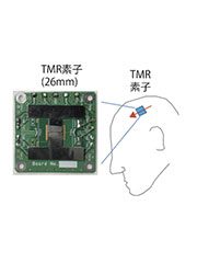
The figure shows the somatosensory evoked magnetic field response waveform measured using the TMR element (upper right) and the waveform measured using the superconducting element (lower right). The two waveforms, presented in similar formats, show that the amplitude captured by the TMR element is higher than that captured by the superconducting element. The upper left panel shows the TMR element.
Future spread of the MEG system that can be used anytime and anywhere
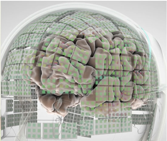
The successful recording of weak magnetoencephalography in the present study opened up the prospects for a general-use type TMR-MEG system(Fig.4). Since the TMR sensor is resistant to external noises, including magnetism generated from surrounding electronic devices, it is expected to enable examination of brain functions in regular daily life outside the special magnetic shielding room for diagnosis of epilepsy, etc. The low manufacturing cost makes mass production possible. In addition, it does not require the cost of liquid helium for cooling, and the compact portable device can also reduce operating costs. Future development and an explosive spread of inexpensive MEG systems that allow measurement to be performed anytime and anywhere are expected to benefit many epileptic patients.
Moreover, the results of the present research will further promote medical application and social implementation of spintronics, which is Tohoku University’s area of expertise.

Fig.4 Future magnetic field measurement using TMR elements
In the future, it is expected to become possible to perform measurements by placing hundreds to thousands of channels in contact with the scalp.
- Life Science
- Research Results
- Japanese
