Research Results
Creation of experimental resources applicable to the development of treatments for diseases
Successful Development of Mice to Observe Cell Proliferation in Live AnimalsFY2024

- KATAGIRI Hideki (Professor, Tohoku University Graduate School of Medicine)
- Moonshot Research and Development Program
- Moonshot Goal 2: Realization of ultra-early disease prediction and intervention by 2050
Project Manager (2020-2025), "Challenge for Eradication of Diabetes and Comorbidities through Understanding and Manipulating Homeostatic Systems"
Development of mice that allow observation of cell proliferation in vivo
A research group led by Hideki Katagiri (Professor of the Department of Metabolism and Diabetes, Tohoku University Graduate School of Medicine, and the Department of Diabetes, Metabolism and Endocrinology, Tohoku University Hospital) has successfully developed mice that allow temporal and highly sensitive observation of the proliferation of cells*1 to be examined in vivo with a very small amount of blood collected.
Previously, observation of cell proliferation over time in the body of a living animal has only been achieved by collecting and analyzing animal organs at multiple time points, requiring substantial experimental resources, including animals.
Using these mice, Professor Katagiri et al. have succeeded in in vivo monitoring on a continuing basis of the proliferation of pancreatic β-cells,*2 which are few in number, in addition to liver cell proliferation in the same mouse. Furthermore, the cells from these mice were found to have potential use in the search for cell-proliferative and anti-proliferative drugs. Because these methods allow tracking of changes over time in the same individuals without being affected by individual differences, they are expected to advance the development of treatments for various diseases, such as curative drugs for diabetes that increase insulin-producing cells and drugs that inhibit cancer cell growth, while making effective use of experimental resources.
*1 Proliferation of cells
Increasing the number of cells by dividing one cell into two. A mechanism that is critical for maintaining organs at their normal function and size. In addition, cancer cell proliferation is a known cause of cancer growth.
*2 β-cells
The only cells in the body that produce insulin, a hormone that lowers blood glucose levels. They are clustered in many islet-like areas in the pancreas known as islets of Langerhans. Diabetes is known to develop when the function of these β-cells is impaired or decreased in number.
Methods for studying β-cell proliferation needed to overcome diabetes
As the project manager of the Moonshot Research and Development Program, Professor Katagiri has pursued research on the subject "Challenge for Eradication of Diabetes and Comorbidities through Understanding and Manipulating Homeostatic Systems," with the aim of detecting and overcoming diabetes and its comorbidities at a very early stage as simply and conveniently as possible. Diabetes is a disease caused by decreased levels and/or impaired function of insulin that lowers blood glucose levels. Decreased insulin levels result from reduced number of insulin-producing β-cells. Searching for substances that increase β-cells is essential for treating diabetes, but until now it has not been possible to study the proliferation of β-cells in vivo.
Therefore, there has been a demand for the development of a simple and convenient method for studying cell proliferation.
Aiming for a high-speed, high-sensitivity, and low-cost automatic detection system
The research group first created genetically modified mice that were designed to produce a protein called luciferase and release it into the bloodstream when proliferation of specific cells occurs. By drawing a small amount of blood from these mice and measuring the amount of luciferase in the blood, it made it possible to detect the proliferation of specific cells occurring in vivo in real-time (Fig. 1). Using this method, it is possible to observe the state of cell proliferation in the same mouse as many times as desired. In addition, by changing the mechanism of genetic modification depending on the type of the intended cells, various types of cell proliferation can be observed.

Fig.1 Genetically modified mice that allow detection of the proliferation of specific cells
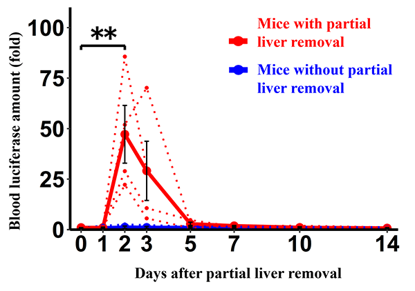
Next, the research group observed the proliferation of liver cells using these mice. It has been demonstrated that when the liver is partially removed, the remaining liver cells proliferate and maintain the size and function of the liver. Therefore, the research group partially removed the livers of these mice and then collected blood samples to observe proliferation of the liver cells over time. As a result, they were able to detect, with extreme sensitivity, changes in the proliferation of liver cells (Fig. 2).

Fig.2 Continuously detected changes in liver cell proliferation after partial liver removal
The research group succeeded in continuously detecting changes in liver cell proliferation in vivo after partial liver removal in mice by drawing a small amount of blood.
They also observed the proliferation of insulin-producing pancreatic β-cells, of which there is a very small number in the body. In previous studies, Professor Katagiri et al. discovered that the proliferation of β-cells is caused by neural signals. So, they stimulated β-cell proliferation in these mice with neural signals and observed the mice over time. As a result, they were able to detect β-cell proliferation (Fig. 3). β-cell proliferation is known to occur during pregnancy, obesity, and the juvenile phase, and these changes over time could also be detected using these mice (Fig. 4). These findings showed that, by using this method, even the proliferation of cells that are small in number can be detected with high sensitivity.

Fig.3 Continuously detected changes in β-cell proliferation
(A) Successful continuous detection of changes in β-cell proliferation in vivo in mice stimulated with neural signals by drawing a small amount of blood
(B) β-cell proliferation detected through this newly developed method strongly correlated with β-cell proliferation detected by the conventional method using pancreatic specimens
**With large significant differences

Fig.4 Continuously detected changes in β-cell proliferation during pregnancy, obesity, and the juvenile phase
(A) Successful continuous detection of changes in β-cell proliferation in vivo in mice during pregnancy by drawing a small amount of blood
(B) Successful continuous detection of changes in β-cell proliferation in vivo in mice during obesity by drawing a small amount of blood
(C) Successful continuous detection of changes in β-cell proliferation in vivo in juvenile mice by drawing a small amount of blood
*With significant differences **With large significant differences
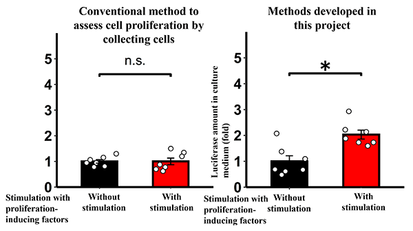
In addition, when these mouse β-cells were subjected in vitro to substances known to induce β-cell proliferation, luciferase in the cell culture medium increased, and cell proliferation was detected with high sensitivity (Fig. 5). These results suggested that the use of this method may help to advance the search for substances that increase β-cells.

Fig.5 Detected β-cell proliferation in vitro by proliferation-inducing factors
Successful detection of β-cell proliferation in vitro by proliferation-inducing factors, which has been difficult to detect through conventional methods of cell proliferation assessment
n.s.: Not significant *Significant
Contributing to the development of methods for the prevention, diagnosis, and treatment of diabetes
Through this research, the group succeeded in developing mice that allow observation of changes over time in the proliferation of the intended cells in vivo with only a very small amount of blood collected. By using these mice, progress is expected in the development of unprecedented therapeutic agents for various diseases, for example, through increasing insulin-producing pancreatic β-cells for diabetes, increasing heart cells for heart failure, and increasing muscle cells in elderly people with reduced muscle mass, while markedly saving experimental resources.
On the other hand, this research is expected to contribute to the development of drugs to suppress cancer by inducing cancer in these mice and observing the proliferation of cancer cells when the drugs are administered.
In the project led by Hideki Katagiri as part of the Moonshot Research and Development Program, the group has been developing methods for prevention, diagnosis, and treatment of diabetes using inter-organ networks via the nervous system. On November 9, 2023, they published an article titled "Optogenetic stimulation of vagal nerves for enhanced glucose-stimulated insulin secretion and β cell proliferation" from Nature Biomedical Engineering as a new research result. This article describes how they succeeded in increasing insulin-producing pancreatic β-cells by stimulating the vagus nerve, a type of autonomic nerve that connects the brain and the pancreas.
- Life Science
- Research Results
- Japanese
