Research Results
Elucidating how the brain works during “empathy”
Discovery of neurons that hold combined information about self and othersFY2024

- OKUYAMA Teruhiro (Associate Professor, Institute for Quantitative Biosciences, The University of Tokyo)
- Fusion Oriented Research for Disruptive Science and Technology (FOREST)
- Elucidation of the representation mechanism of “self” and “others” in the brain (2022- up to 10 years)
Discover how the brain works when empathy occurs
Both humans and mice are known to “empathize” with feeling of fear themselves when they see others experiencing fear, as if those feelings are transferred to them. However, the detailed neural mechanism behind this has not yet been elucidated.
In this study, Teruhiro Okuyama, Associate Professor at the Institute for Quantitative Biosciences, The University of Tokyo, and his research group used mice to elucidate how “others’ emotions” and “one’s own emotions” are processed in the brain when one feels fear upon seeing others feeling fearful.
At this time, the research group discovered that in the brain region called the prefrontal cortex, there are neurons that simultaneously hold and express combined information about one’s own and others’ emotions. These findings are expected to advance our understanding of autism spectrum disorder, which presents difficulties with empathy.
Explore the neural mechanisms of emotional contagion
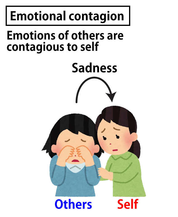
The phenomenon in which emotions are “contagious” from one person to another, such as “when I see my best friend feeling sad, I feel sad as if it were me,” is called emotional contagion (Fig. 1).
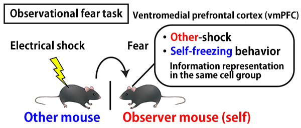
Since this phenomenon is observed not only in humans but also in many animal species, such as mice, research has been conducted to unravel its neural mechanisms through “observational fear behavior” using animals. In the observational fear behavior, it can be confirmed that the observer mouse also displayed a fear response upon witnessing another mouse receiving electric shocks and exhibiting a fear response (Fig. 2). Previous studies have focused on the “freezing behavior,” in which mice cower and tremble on the spot as a fear response in the observer mice, and analyzed the neural mechanisms at that moment. The results showed that brain regions such as the anterior cingulate cortex (ACC), which is involved in pain perception, and the basolateral amygdala (BLA), which controls emotion, are involved in freezing behavior. However, the observer mice exhibited a variety of behaviors other than the freezing behavior, and the neural mechanisms of these behaviors remained unclear.
In this study, the research group analyzed the function and neurophysiological characteristics of the ventromedial prefrontal cortex (vmPFC) in observational fear behavior, which previous studies have suggested may play an important function in the process of observing and empathizing with fear in humans, and further investigated the role of neural inputs from the ACC and BLA to the vmPFC.

Fig.1 Emotional contagion

Fig.2 Observational fear behavior experiment
Analysis of various behaviors of observer mice using automatic classification method
By combining deep learning-based technology for tracking animal body points with dimension reduction clustering*1, the research group has succeeded in objectively and automatically classifying the complex behavioral patterns exhibited by the observer mice during observational fear behavior.
This automatic behavior classification method was used to analyze various behaviors of the observer mice. When optogenetic inhibition*2 was applied to the vmPFC in the observer mice, it was found that although “freezing behavior” was not reduced, behavioral changes such as “gazing behavior toward others undergoing fear” increased and “escape behavior” decreased. Optogenetic inhibition of neural inputs from the ACC and BLA to the vmPFC, respectively, increased escape behavior, contrary to when only the vmPFC was inhibited. These analyses indicate that the vmPFC and the neural inputs of ACC→vmPFC and BLA→vmPFC are mainly involved in the control of “escape behavior” during observational fear behavior.
Then, in order to examine the information held by the neurons in the vmPFC of the observer mice, “calcium imaging*3 using brain microendoscopy” was conducted to observe the neural activity in the brain during the observational fear behavior. As a result, it was found that the neurons reflecting specific behavioral states of the observer mice were present in the vmPFC. Furthermore, the behaviors exhibited by the observer mice could be decoded*4 from the activity of the neuronal population, indicating that the neurons in the vmPFC have information on behavioral states of the self.
Additionally, it was found that the vmPFC also contains neurons that respond to electric shocks in other mice. Interestingly, it was revealed that the group of cells with information about other-shock overlapped with the group of cells with information about self-freezing behavior (Fig. 2). This suggests that there are neurons in the vmPFC that “simultaneously” hold combined information about both “self-fear” and “other-fear”. Moreover, when the neural activity of the vmPFC was recorded in a similar manner while optogenetically inhibiting neural input from the ACC and BLA, anomalies were found in the characteristic of “simultaneously” hold combined information about both “self-fear” and “other-fear.” The results suggest that the information input from the ACC and BLA to the vmPFC is both due to the information processing of one’s own and others’ emotions in the vmPFC.
*1 Dimension reduction clustering
In this study, dimensional reduction, a method of reducing high-dimensional behavioral data to a lower dimension without losing as much information as possible, was first performed. After drawing the data on a two-dimensional plane, the data points were grouped (clustered) according to the distribution density of data points on the plane using the watershed algorithm.
*2 Optogenetic inhibition
In optogenetics, a photoreceptor protein that is activated by light of a specific wavelength is first expressed in a specific group of cells using genetic techniques. This group of cells can then be excited or inhibited by light illumination only in specific periods, such as during behavioral experiments.
*3 Calcium imaging
A method of measuring intracellular calcium flow using microscopic techniques. Because calcium ion concentrations in neurons change with neural activity, neuronal activity can be recorded by measuring the amount of fluorescence of calcium indicators, whose fluorescence intensity changes in a calcium concentration-dependent manner.
*4 Decoding
Readout and estimation of external stimuli, behavioral and cognitive states, etc., from measured neuronal activity.
Expectations for application to autism spectrum disorder with difficulties in empathy
Empathy is a phenomenon in which the boundary between “self” and “others” seems to temporarily disappear, and plays an important role in building good communication in our daily lives. The neurons found in this study, which hold combined information about both “self” and “others” during the occurrence of empathy, are thought to contribute to a brain mechanism that makes the boundary between self and others feel like it is disappearing.
Associate Professor Okuyama and his colleagues have been conducting research to elucidate the pathophysiology of autism spectrum disorder, which is characterized by ambiguous boundaries between self and others and difficulties in empathy toward others’ feelings. The results of this study are expected to advance our understanding of autism spectrum disorder.
- Life Science
- Research Results
- Japanese
