Research Results
Transcending the limitations of conventional X-ray imaging devices
Contributing early diagnoses with a Super X-ray FY2016
- Atsushi Momose (Professor,Tohoku University) / Konica Minolta,Inc.
- Development of Advanced Measurement and Analysis Systems
- Technology development (2004-2007), System development (2007-2011)
Practical Realization (2011-2013), Promotion for common use of outputs (2011-2013)
Developing an X-ray grating interferometer apparatus -Development of a diagnostic devise based on differential phase contrast by X-ray Talbot-Lau interferometry-; Team leader
Scanning soft tissue as well!
Developing a revolutionary new X-ray apparatus
In conventional X-ray imaging used for bone examinations and so on, contrasting shades are obtained depending on the extent to which the object absorbs X-rays. When examining tissues such as organs, for example, since contrasting shades cannot be sufficiently obtained with this method, X-ray photography is conducted after introducing a contrasting agent such as barium into the body; alternatively, advanced scanning technology such as the MRI must be used. If some method could be developed to use X-rays to provide images of soft tissue, this would lighten the patient burden, and would also likely prove useful in the early detection of diseases.
For the last 20 years, Professor Atsushi Momose has been working on development of just such an X-ray imaging apparatus. In order to develop an apparatus useful for medical practice that transcends the limitations of conventional X-ray imaging devices, Professor Momose has been conducting various kinds of research.
This X-ray photography method, based on a completely new principle discovered by Prof Momose, is currently being developed for commercialization by medical equipment manufacturer Konica Minolta. It has already been successfully used for early detection of breast cancer lesions and imaging of cartilage, and it is expected to bring further benefits to the front line of medical practice.
Conventional method (Absorption image)
Differential phase image
Scattered image
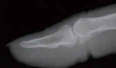
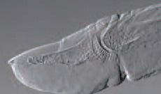
A real radiography image of thumb took by a X-ray photography apparatus has succeeded in developing (middle: differential phase image, right: scattered image).
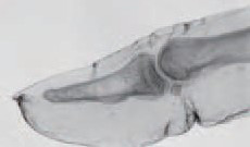
Tissues such as cartilage and tendons were taken by its apparatus use the traditional method.
Using slight X-ray refractions to enable the scanning of soft tissue
In 1836, the British scientist Henry Fox Talbot discovered the phenomenon known as the "Talbot effect," whereby waves of light passing through a substance with a regular structure such as a grating form selfimages at specific distances. Just as with light, X-rays possess wave properties. By applying the Talbot effect (brought to light almost 180 years previously) to X-rays, Professor Momose believed he could realize a new high-precision imaging technology.
In the early 1990s, before becoming interested in the Talbot effect, Professor Momose detected X-ray refraction with an apparatus known as an X-ray interferometer using silicon crystals, thus proving that high sensitivity X-ray photography was possible. Using X-rays, this method was able to provide images of cancer tissue; however, it required a massive source of special X-rays and so was not adopted for immediate medical application. In the 2000s, Professor Momose started developing a "Talbot interferometer" using the Talbot effect, achieved by placing two gratings between the X-ray detection device and the object scanned. With the Talbot interferometer, X-rays pass from the X-ray source through the object and then pass through the first grating. Since the speed of X-rays passing through the solid object changes, the wave's position goes out of alignment and refraction occurs at around a ten-thousandth of 1 degree. As a result, the self-image that appears due to the Talbot effect is distorted. Subsequently, by placing a second grating corresponding to this self image, differences in the extent of X-ray penetration are created, depending on the distortion, which forms a moiré pattern that can be observed. With analysis of this pattern, Professor Momose has successfully established a method for the imaging of soft tissue such as internal organs.
In 2004, as part of the Development of Advanced Measurement and Analysis Systems,, development of X-ray imaging apparatus using the Talbot effect commenced.
In order to obtain the Talbot effect, it is necessary to form the X-rays into waves. In initial development, a light source capable of emitting X-ray formed in waves (a micro-focus X-ray source) was used, with practical use in hospitals expected. However, the X-rays emitted from the light source were weak and sufficiently good images could not be obtained given practical imaging times.
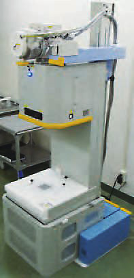
Prototype X-ray photography apparatus (vertical model) (Provided by, Konica Minolta Inc.)
Utilizing the X-ray used in hospitals!
The X-rays used in hospitals are not formed into waves, which mean they cannot use the Talbot effect unmodified. This problem was solved by installing one more grating close to the general X-ray source. The newly added grating is designed to only allow X-rays formed into waves to pass through to the Talbot interferometer. With this solution, X-ray sources that have been successfully used in hospitals can be utilized for X-ray photography utilizing the Talbot effect; thus, the Talbot low X-ray interferometer was born, which also effectively cuts down photography time.
This revolutionary X-ray photography apparatus is capable of imaging of tissue with early stage breast cancer, soft cartilage and other conditions that are difficult to image with conventional methods, and it is expected to bring great results in medical practice. Furthermore, with high spatial resolution and online testing equipment practical development of the three-dimensional imaging for the realization of the X-ray non-destructive testing for the possible precision testing equipment has begun. This device expands to use not only medical field also used for the purpose of safety and security and industrial production management such as organic materials and devices.
Composition of X-ray Talbot-Lau interferometer
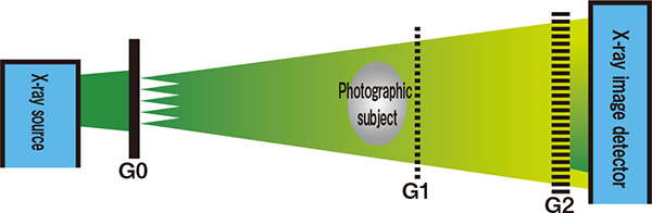
Using 2 plates (G1andG2) installed another plate close to close to light source of the X-ray Talbot-Law interferometer. It is possible to extract only x-ray coherent even the x ray-sourse used in the hospitals.
Flexible thinking brings about revolutionary results
"Researchers need to have a playful mentality. The way you use your brain when conducting research is more like arts and crafts or music than undergraduate study,"says Professor Momose. By incorporating flexible thinking with the Talbot effect, a 180-year old concept which had not been utilized in the world of X-ray technology, a new technology has been successfully created. The revolutionary X-ray photograph apparatus developed in this way is expected to contribute greatly to the early detection of diseases.