Developmental simulation of a musculoskeletal and nervous system.
Fetuses and Infants musculoskeletal nervous system simulation.
Construction of fetal and infants’ model simulation environment
To reveal the mechanism of human cognitive development,
we decided to construct a baby computer simulation system at the beginning of the project.
Human infants are confirmed already to have kinds of cognitive functionality at the birth by behavioral experiments and observations,
however we do not know why and how they acquire the ability without experiences after birth.
This is the big issue “Nature or Nurture”
To study the mechanism of human development,
it is necessary that we investigate developmental process before and after the birth.
We need computer simulation model of human fetuses and infants
so that we study the principle mechanism of cognitive development from the perspective of embodied cognition.

Fig.1 Body organization of fetuses and infants models.


Fig.2 A free form fetus model.

Fig.3 An example of behavior of a free form infant model.
First of all, we constructed fetuses and infants model by arranging spheres and cylinders (Fig.1)
[Kuniyoshi2006].
The model parameters are based on literatures and our intensive investigation in some medical and physiological institute (Tokyo Women’s Medical Universtiy, RIKEN and so on).
The models has 198 muscles and sensors, which are muscle spindles, muscle tendon and vision.
After the construction, we constructed free form models of fetuses and infants to generate realistic tactile sensation.
The models has over 1500 tactile detecting points and the distribution of the points can be changed to investigate the effect of sensor conditions to behavioral develpment.
We made generator program of fetuses and infants align with desired age and parameters are decided depending on the age.
This kind of simulation system is very unique and has no precedent in the world.
The models have been improved by group members actively and used for various research issues.
We have achieved many important results about behavioral and neural development of fetuses and infants from the perspective of behavior differentiation from general movements generated by spinobulbar neural oscillators.
We report the results as follows.
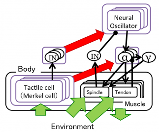
We proposed a hypothesis of a mechanism of early fetal behavioral developments.
The hypothesis is that tactile sensations induces the behavioral development.
So we conducted simulated experiments with human-like tactile distribution and artificial uniform distribution,
By the experiments, we confirmed that human-like tactile distribution induces human-like behavioral developmental process
[Mori2010].

Fig.4 Primitive nervous system model with spinal cord and medula.
Fig.5 Appearance of a fetal simulation.
We constructed a nervous system model for late fetal period with primary somatic and motor cortex and conducted a developmental experiment[Kuniyoshi2006].
The result shows self-organization of somatotopic map in the cerebral cortex and the behavior simplified through an experience in utero.

Fig.6 Nervous system model with spinobulbar system and primary somatic and motor cortex.
Fig.7 infant’s model and emergent rolling over movement and crawling movements.
We constructed a cerebellum model to memorize and reuse explored behaviors by spinobulbar system.
In the experiment, behaviors can be memorized in cerebellum model without value function,
and repeated for composite behaviors
[Kinjo2008].
We proposed a hypothesis of self-organization of goal-directed movements induced by sensory constraints.
We interpreted that fetal hand and face contact behaviors are induced by dense tactile cells in hands and a face,
and infant’s hand regard behaviors are induced by the constraint of the visual area.
So we constructed nervous system model (Fig.8) and conducted fetal and infant’s simulation.
As the result, hand and face contacts and hand regards increase only with sensory constraints (Fig.9)
[Yamada2010].

Fig.8 Spinobulbar and primary somatic and motor cortex with a neuromodulator model.

Fig.9 An appearance of hand regards experiment by a infant’s model.
We investigated relationship between body and spacial representation, from perspective of theta rhymes in hippocampus, and reaching.
A EEG research with 2 to 11 months old infants indicated that theta rhythms, which has relation to hipocampus, become active in Parietal lobe and Premotor area at a reaching and a thumb-sucking
From the insight, we hypothesize that Hippocampus induces infants’ preference to movements related to themselves and the preference affect reaching and spacial representation in the brain.
Proposed spiking hippocampus model with theta coding became to present spaciotemporal relationship by STDP learning rule,
and reaching behaviors increase along with learning (Fig.10)
[Pitti2010].

Fig. 10 Learning by hippocampus model. Sensory information synchronized to theta wave (6Hz) are transformed to spiking signal in hippocampus (Left fig.), Learning occurs depending on spaciotemporal spiking synchronization.
