Research Results
Visualize cancer cells by simple spraying
A light of hope for the improvement of residual breast cancer after surgery FY2016
- Yasuteru Urano (Professor at the Graduate School of Pharmaceutical Sciences and Graduate School of Medicine, The University of Tokyo)
- PRESTO
- Structure Function and Measurement Analysis "Development of advanced photo-functional probe molecules for elucidation of biological events in the living cells" Researcher (2004-2008)
- Research Acceleration
- "In vivo tiny tumor detection project using light functional probe" Research Director (2010-2015)
- CREST
- Creation of Innovative Technology for Medical Applications Based on the Global Analyses and Regulation of Disease-Related Metabolites "Creation of search techniques for disease-related metabolic activities based on live imaging of clinical specimen and its application to drug developments" Research Director (2014)*
*This project was transferred to the Japan Agency for Medical Research and Development from fiscal 2015.
Development of the world's first fluorescent reagent that enables extremely rapid detection of cancer site
Early discovery and treatment is important in cancer. Prognosis can be improved dramatically if the primary lesion is removed before metastasis, and if the metastatic tiny tumor can be removed even after metastasis. In recent years, the treatment methods are shifting towards endoscopic or laparoscopic surgery to reduce the burden, creating a high demand for the establishment of methods that enable accurate detection of cancer location.
The limit of cancer detection is around 1 cm with the present method of cancer screening, and it is extremely difficult to detect cancer with the size of a few millimeters. In order to prevent the recurrence of cancer, there is a need to remove all metastatic tiny tumor around 1 mm in size, however this was previously dependent on the experience of the operating surgeon, and failure in detection and removal was an issue.
In Research Acceleration, Professor Yasuteru Urano and principal investigator Hisataka Kobayashi of the National Institutes of Health (US) conducted a joint research and succeeded in a development of an innovational reagent which enables only cancer cells to be detected in a short period of time. By simply spraying a small amount of "organic small molecule fluorescent probe", a pigment that visualizes the substances in the living body, onto the affected area, fluorescence appears only in the area of the cancer. This is the first technology in the world which enables the area of cancer to emit visible fluorescence by spraying locally, and it is a truly innovative technology.
Fluorescence detection image of the cancer sites
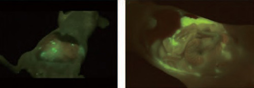
By spraying the probe reagent, the cancer sites emit a strong fluorescence after about 1 minute. It is possible to detect the area of tiny tumor by visual inspection (mouse model with peritoneal metastasis of ovarian cancer).
Tiny tumor at the size of 1 mm or smaller can be detected in a few tens of seconds to few minutes
In this research, Professor Urano and his colleagues focused on "γ-glutamyl transpeptidase (GGT)", a protease whose activity is increased in many cancer cells, such as ovarian and lung cancer. Based on the original fluorescent probe design established by the professor himself, the world's first fluorescent imaging reagent was successfully developed that detects the GGT activity of living cells as green fluorescence. This reagent itself is colorless and dose not emit fluorescence, however it changes into molecules which emit strong green fluorescence when it comes in contact with GGT. This enables fluorescent staining only for cells that have high GGT activity in a short period of time.
This reagent was tested in model mice, which revealed that tiny tumor at the size of 1 mm or smaller can be detected clearly in a few tens of seconds to a few minutes after spraying in the area of the suspected cancer. The strong fluorescence at the cancer site could be observed not only with a fluorescent endoscope but also by visual inspection. In addition, this reagent visualized the undetected very tiny tumor areas in the body of the model mice and succeeded in the mock surgery of removing these tumors with forceps.
Effective in the actual surgery for breast cancer
The effectiveness of the developed fluorescent imaging reagent was demonstrated in cancer model mice. However, the nature of cancer in human is highly diverse, and it was not known if the reagent would be effective for actual surgery. In a joint research with Professor Koshi Mimori of Kyushu University Beppu Hospital, Professor Urano has attempted to confirm efficacy in human cancer samples. As a result, spraying of this reagent on the clinical sample resected in breast surgery has shown strong fluorescence in tiny tumor at the size of 1 mm or smaller after about 1 to 5 minutes of spraying, enabling clear distinction of breast cancer from the surrounding health tissues such as mammary glands and fat. This method revealed that 1 mm or smaller size cancer tissues in small mammary ducts can be detected.
In the actual partial mastectomy of breast cancer, there is a need to detect small lesions (1 mm or smaller) in a sample the size of a few centimeters. In order to check whether the cancer cells have been completely removed, the cut end of the resected specimen must be checked for cancer cells during the surgery. However, in the present days where there is a shortage of pathologists, it is difficult to test all specimens, and the remaining cancer may be let undetected. This reagent is considered innovative in that it eliminates failure of detection in pathological diagnosis, thereby preventing the failure to remove cancer in surgery.
Detection of small cancer in the surgery specimen using the fluorescent reagent
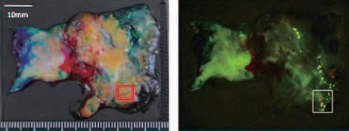
In the large sample at the size of about 5 cm (left diagram) contains small hidden cancer in the area shown with a red rectangle in the right bottom corner. Although this cannot be seen with the naked eye, a few areas of strong fluorescence were detected about 5 minutes after spraying the reagent (right diagram). This section was subject to detailed pathological examinations.
Magnified diagram of the photograph above and the result of histopathological staining
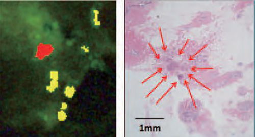
Success in detecting small cancer around 1 mm in size, which cannot be detected visually
(The areas with red and yellow in the left diagram shows strong fluorescence. Of these, the red area was proved by histopathological staining to be identical to the area of cancer in the right diagram).
Clinical evaluation conducted by industry-academia collaboration towards commercialization
Because of the rapid and convenient nature of fluorescent detection and the affordable cost, this technology has a high expectation for being used widely as a general method of cancer detection. Currently, clinical evaluation is conducted by industry- academia collaboration to investigate the safety and efficacy of the reagent with the aim of commercialization as clinical pharmaceutical. However, there are types of cancer that have almost no expression of GGT used as a target in this reagent (some cases of colon cancer, ovarian cancer, and gastric cancer), and the development of new reagents that visualize cancer tissues in these types of cancer is now on-going. It is expected that in future most cancer sites will be detected in a short period of time. In any case, development of this reagent is likely to be good news for many patients with cancer, and a brings dramatic change to the future of cancer as a disorder.